Many claims are being made about what one can do with Live Blood Analysis and this course will blow the trumpet of caution on several popular assumptions. That way you are going to end up with 1) a balanced view and 2) greater clinical confidence. By examining this topic in an comparative way from several angles you will get an excellent grasp of what is reasonable and above all what works in clinical practice!!
If humans are in his feeling as in his thought world in the imbalance, also the acid Basen household is not any more in the equilibrium.
Nutrition, which overacidifies: animal proteins, pointing flour, sugar, coffee, black dte, alcohol, nicotine; evenly all luxuries.
We made our luxuries our food. Since all luxuries overacidify, the accordingly sour environment is prepared for the microbes. One can call many illnesses therefore civilization illnesses. In addition comes frequently more negatively, more interhumanly, more privately like vocational stress. In addition feed lack of movement, electrical smog, environmental contaminations and water veins the sour environment.
Microbe development after of Brehmer: If itself the blood pH value within the ideal range of 7,2 - 7.3 finds, no microbe can develop further into the Pathogenitaet themselves in the environment and to spread. Only if the blood pH value changes into Alkaline from 7,4, seem themselves spontaneously from food proteins an infectious Dns rns (storage mediums of the cell) after so far not well-known regularities, either to sporen, Viroiden, viruses, bacteria or mushrooms develop further. In the dark field such development stages can be pursued.
The health is carried by 4 columns:
the acid Base household
the water regime
the electrolyte household
the respiration; speak oxygen admission
If one of the 4 columns comes into the imbalance, everything comes from the equilibrium; Illness takes its way!!!
It applies to consider: as so in the SMALL ONE in the LARGE ONE.
Most loading factor for the Ur germ, or also the Endobiont or also Protit, is before-all-hires wrong the way of life and thinking, so that the Apathogen can to develop to the Pathogen and be able diseased disturbances to be released. From friends to enemies. The naturopathy is anxious to let from enemies again friends become. In addition the modulators are used.
A most important condition: the patient must help!!!
The Cyclogenie kept until today despite neglect by the training medicine persistent alive. Today she celebrates a considerable national due to the rapid development of the dark field diagnostics and therapy like international Renaissance.
Micro organisms can develop in the Erythrozyten or from the sources of infection devitale teeth (dead teeth), dental herd without life, root granulome (inflammatory fabric) and intestine to develop, of where from the blood is infected over well-known haematogen and lymphogen ways. Both is supposed the case. If the intestine with purposeful cleaning cures and different Therapeutica is reorganized and afterwards by sporen, viruses, bacteria and mushrooms is free, the blood values improve spontaneously!!!
Nevertheless still another further, in the plasma, must be accepted free germ development, as Enderlein , from Brehmer and Hafeli to have always stated. But the blood pH value is responsible primarily.
A) the Mucor Racemosus Fresen and b) the Aspergillus the Niger van Thieghem; in all their stages from the virus to the mushroom. The appropriate bacteria forms of these Cyklod are A) Leptotrichia buccalis Robin and b) Mycobakterium tuberculosis.
In the Chondrit area this as physiological and innocuous, even useful, Symbionten in the blood and fabric of healthy humans live. As soon as the biochemical equilibrium of humans changes however, the Chondrite ascends in the higher phases and/or valences and accepts thereby a pathogen character. This applies to some civilization-dependent diseases. Recovery to lower valences takes place only on sexual way via nuclear fusion with sufficiently existing apathogen Chondriten. This procedure is blocked with ill humans. Prof.Dr. Enderlein created for this purpose the Chondritpraeparat Chondritin, with which a recovery of the pathogen valences running as nuclear chain reaction is introduced. The Erythrocyte, normal way memory of the resting Symbionts, accepts the so-called Stechapfelform in these cases frequently.
Cyclogenie means the transformationand migration of all pathogen and not pathogen germs by all phases (valences) of the border of the visibility, and among them the virus range, over the higher valence phases of the text book-in accordance with-eaten Kokken and the staebchen up to the kulminanten phases of the mushrooms and their Myzelien.
The bacteria core (Mych) plays an important role, the moreover the pH value, i.e. the acid Base household of the organism of humans (to the comparison: as is the case for the aquarium). Only if the environment is not correct, the pathogen development can take place. Either asexual by division or on sexual way by Sprouting after preceding nuclear fusion. The principle of the Polymorphic was confirmed 40 years after Enderlein by the Nobel leather castle.
Method of Professor Dr. Guenther Enderlein (1872-1968), that primarily a biologist and a zoologist was 1916 came from Enderlein a first report over a revolutionary reorganization of the bacteriology.
Dr. William of Brehmer (1883 - 1958) discovered 1928 the blood parasites Siphonosphora polymorpha in the red blood corpuscle of humans, which can develop itself further under certain conditions to diseased forms. The special at of Brehmer the method is coloring native or vital blood on a slide, whereby certain high valence forms become visible. By the advancement of the technology to the phase contrast microscope, it is to be regarded us today possible certain forms without coloring immediately.
Most important aspect also for of Brehmer is clarifying the acid Base environment in the organism. It developed for it the Haemo Ionometer. Robert-Koch-Institute with the Humboldt university in Berlin, under which line of the then world-famous Hematology Professor Dr. Victor Schilling confirmed to 1935 that Siphonosphora polymorpha the second genuine blood parasite is, which develops in the Erythrocyte. First up to then admitted blood parasite was the Plasmodium malaria. Thus Brehmers discovery was scientifically certified and in the medical professional world recognition.
The exciters go through a specific cycle, as the training medicine and bacteriology accept it with malaria as natural. The training medicine until today recognizes the development stages of viruses, bacteria and mushroom forms however not on. Although there is nevertheless no exception of the law of the eternal change and the unit of the macrocosm with the microcosmic (e.g. Qualquappe' Frog, Raupe' Butterfly) in whole nature.
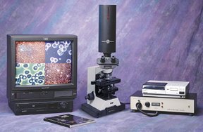
Live Blood Analysis uses a drop of live blood from the patient's finger that has not been killed by staining, and viewed under a special microscope using a darkfield condenser.
This enables the blood sample to be illuminated from the sides, making the various components phosphoresce behind a dark background.
This makes it possible to see very small particles, smaller than a cell that would not normally be visible under a normal light microscope. All the living components of the blood are seen clearly, and can be viewed by the patient and therapist using a video camera and a dedicated monitor.
The examination of live blood is valuable for the early detection of serious health conditions. It is possible to see at what stage of pathological development the body is in, simply from using one drop of blood.
Because the precursors to serious health imbalances may be observed in the state of ever-present floras found in the blood, health imbalances may be averted by reading these early warning signs and making the necessary changes that will allow one to rebalance the physiology. These markers are also applicable in the course of tracking the progression and reversal of degenerative conditions that may already be in motion.
DARKFIELD MICROSCOPY The observations of these floras are made using what is known as a Darkfield microscope. When utilizing today's conventional approaches to analyze blood, the standard methods are to use either stains, which make certain factors in the blood visible which would not be otherwise visible, or the electron microscope, which provides ultrahigh magnifications. The limitation of both of the preceding approaches is that the blood is effectively killed through the processes utilized in observation.
In a Darkfield, the blood is stained with light frequencies, which allow the blood to remain alive and active, giving many otherwise unobtainable clues as to the relative health of the blood, thereby, the whole organism.We have entered into a new era of scientific discovery. As the horse gave way to the horseless carriage, so must science now accommodate a new understanding of the body as a whole.
Edgar Cayce, the seer of Virginia Beach, predicted in the 1930's that in the future, a person's state of health would be determined by the evaluation of one drop of blood. This time has arrived!Through the advent of technological advances in microscopy, new discoveries have been proven by such leading researchers as Royal Rife, Gaston Naessans, Dr. Gunther Enderlein, Dr. Majid Ali, M.D. and many others. In the rapidly emerging field of what is known as live cell analysis, an understanding of biology as a holistic science has emerged. Health imbalances in the body may be averted by observing the state of ever-present floras found in the blood and by correcting the milieu that allows the floras to remain in their regulatory forms or to move into pathogenicity. Or conversely, to be reduced from pathogenic forms back down to regulators. These observations are made in what is known as a darkfield.
DARK-FIELD SYSTEMS Under dark-field examination, the various materials making up the structure of the cell or microorganism under view cause it to appear to glow and emit its own light. Since fine structures often cannot be seen when they appear in front of a bright background, illuminating them from the side and viewing them on a black background lights them up and provides contrast and super fine resolution when plan achromat oil immersion iris diaphragm objectives are used. The background is black because the direct or transmitted light from the condenser is passing around the objective and not directly into it. The only light that is seen is light that is reflected off of the sides and surfaces of the particles. What are seen are illuminated particles on a black background. This provides a very clear view of the minutest forms, as the eye's potential to differentiate is not overwhelmed by background light and light passing through the sample. Viewing blood in a dark-field is a very useful adjunct when evaluating and addressing the traumatic factors in the total life picture of the individual. The advanced phases of the life-cycle of the colloidal microorganisms are at the very deepest level of causes of blood imbalance and other organism aberrations .
WHAT CAN BE DETERMINED FROM LIVE BLOOD ANALYSIS? Depending on the irregularities found in the blood, there are a wide variety of different conditions that can be determined. The following are just a few examples:· Indication of low immune status· Liver and spleen stress· Vitamin and mineral deficiencies· Hormonal imbalances· Fungal infections· Parasite infestation· Digestive problems· Atherosclerotic predisposition· Heavy metal toxicity· Predisposition to cancer or other degenerative diseases
IS BLOOD STERILE? For a long time many researchers and doctors believed, and many still do, that blood is sterile. Professor Dr. Gunter Enderlein after numerous years of research, proved otherwise. He showed using darkfield microscopy, that the serum of all people and warm-blooded animals are alive with many moving 'particles'. He called these particles ENDOBIONTS (from the Greek "endon" = internal and "bios" = life). When you first look at a live blood sample under a microscope or on a monitor you cannot fail to see a multitude of moving, living particles. These are the Endobionts, and are an important part of health.
There are a wide variety of different Endobionts in the blood. In fact, from the simplest apathogenic (non-disease forming) PROTIT, they can change forms into pathological (disease causing) species, depending on how much the pH (acidity-alkalinity) of the blood is changed. The higher the acidity, the more pathogenicity, and the more likelihood of developing a chronic, degenerative disease process over time. The wonderful world of Live Blood Analysis can enable you to see the different forms in the blood, and to therefore determine how far ahead you are in the disease process.
Prof. Enderlein discovered that these Endobionts change into different forms as the internal milieu changes. The more the metamorphosis (different changes), the greater the probability of disease. The pH of our blood is determined by the food we eat, and the nutrients we take in daily, as well as other factors such as stress and pollutants. Fast and refined foods and concentrated protein foods all lead to acidic blood which triggers the Endobiont to change to more pathogenic forms. All microbes partake in a natural developmental cycle, that begins with the PRIMITIVE PHASE which is microscopically invisible; this changes into the BACTERIAL PHASE; and finally culminates in the FUNGAL PHASE, which is the most pathogenic stage. This is the theory of PLEOMORPHISM (many forms), as opposed to monomorphism (one form).
This is a 3-day very intensive course with Dr. Eddy Discussion of therapeutic intervention will include antihomotoxic remedies, nutritional & dietary intervention, herbs, glandulars, well-researched enzyme preparations and also some remedies. Next to the copious course notes, each attendee will receive a complementary Biological Analysis script which is another, yet very related course in it own right. This gives the video microscopy course a very broad footing indeed. If you were looking for worthwhile training on a worthwhile topic, then you have found what you were looking for.
3 Days Course Coverage Friday with Dr. Eddy 10 am - 6 pm 1hr lunch
brief introduction of course attendees
concepts: Integrative Medical Clinic, Colon Cleansing, Ozone Treatment, Why Ozone?
medical physiology appropriate to the Milieu Intérieur
Darkfield and Phase Contrast optical physics - Internet links
hematology, starts with normal cell morphology and function, basics hematology
pleomorphism I: Bacteria Cyclogeny and Somatidian Cycle, Protit, Endobiont
Cycle of Imbalance, Cycle of Balance
setting up your microscope & how to take care of it
practicum I: microscopes & Live Blood
Saturday with Dr. Eddy9am - 5 or 6 pm 1hr lunch
confidently recognizing what's under the microscope; abnormal cell morphology of red blood cells, white blood cells, thrombocytes and serum issues including bacterial forms, mycoplasma, crystalline deposits, ascits, filits, fungus/yeast markers, heterogenous plaque etcetera
physiology behind the OST/MOST/HLB™ test i.e. the dried layered blood analysis test
appropriate OST/MOST/HLB™ test pattern recognition
medical physiology & pathology relating to the anomalies you may see
practicum IV pertaining particularly to the OST/MOST/HLB™ test
more general clinical cleansing management
appropriate recapitulation of Friday and Saturday material
blood images quiz to strengthen your gathered knowledge
certificate of attendance signed by tutor (s)
Course notes consist of full color note-page-printouts of some 300 PowerPoint slides. Thus you can follow the tutor (s) slide-by-slide during the course, as well as recall the lecture more easily when you review the material at home. Other than perhaps the odd scribble you need not touch a pen so you can totally concentrate on what is being said & done.
This course is very firmly aimed at enabling health professionals to practice this interesting topic in a clinically responsible fashion. Many claims are being made about what one can do with Live Blood Analysis and this course will blow the trumpet of caution on several popular assumptions.
That way you are going to end up with 1) a balanced view and 2) greater clinical confidence. By examining this topic in an comparative way from several angles you will get an excellent grasp of what is reasonable and above all what works in clinical practice!! Evidence-based Medicine is fashionable so it seemed reasonable to give you an evidence-based course as much as this is possible.
For instance, some courses mention that target cells are linked to a lecithin cholesterol acyl transferase (LCAT) deficiency but few deliver the (patho) physiological model behind it.
After the known enzymatic coagulation cascade and the metabolic/nutritional details to do with it, blood coagulation morphology remains pure pattern recognition. Unfortunately this must be accepted. This course endeavors to explain medical certainties behind the patterns wherever this is possible and discuss differing points of view.
Of course this is a practical course; the day after you should be able to make a good job of testing your family & friends and soon feel confident to use it in your clinic. Following this course you are welcome to e-mail me queries (possibly accompanied by captured live blood images) about a difficult clinical case or microscopes. My one word of warning is do not buy a cheap microscope, they are cheap for good reason. If your field is Optimal Nutrition then why go for equipment that doesn't show you all you need to see to do your job right. Think about that.
Thailand: Integrative Medical Clinic. This Live Blood Microscopy Course costs THB 28,000 for 3 very full days of tuition and includes 1 DrEddyClinic.com Analysis script (which is a related course in itself) and the copious course notes.
The number of attendees is limited to a maximum of 5.
In Thailand, where significantly less people have enrolled for this course as compared to the Continent, shepherding of attendees needs to be done to arrive at an economically viable course i.e. dates are set after a critical number of people has been reached. Not all blood microscopy courses are equal.
It matters hugely what course you take as to what clinical perspective & prescriptions you end up using afterwards. Courses biased towards pleomorphism (Prof. Enderlein) invariably make you biased to practice along those lines. That is why this course covers all aspects, in depth and without bias.
Simply enroll and send an e-mail to Dr. Eddy stating that you want to attend this unique course on Live Blood & Coagulation Morphology Microscopy. He will then e-mail you back for your details and discuss forthcoming Thailand courses. Enrolment is on a strict first-paid first-served basis.
Prof. Enderlein's observations in Darkfield are meticulously documented in his book Bacteria Cyclogeny but there are no (referenced) articles in peer reviewed medical journals available. People have asked if there is any pre-course reading they can do. There are many books on hematology that can refresh your memory on the different blood corpuscules. You must remember that such books predominantly show stained preps and not 'live blood' as we do, but still it is a sensible start.
Books on pleomorphism aren't quite so readily available but you can browse the Internet for the magazine Explore website which will provide you with some interesting pleomorphic articles. Also it may be useful to read the various clinical examples. For now, we look forward to welcoming you on our course.
Further you have here our program, which we providing in all courses. depended which course you choose, you will find different intensity in the study subjects.
 Robert Rowen MD Has Discovered Live Cell Therapies Help Your Body Heal