General Information - The growing utility and popularity of confocal microscopy has resulted in a dramatic increase in applications during recent years and, correspondingly, so have the number of sites on the Internet that address the subject. The links below constitute a selected collection from the best websites available today that provide general information about the history, theory, and practice of this exciting technique. Microscopists at all levels, from beginner to professional, may benefit from visiting many of these resources.
Antibodies - Antibodies are a large family of glycoproteins produced by specialized cells of the vertebrate immune system in response to foreign molecules (antigens). The interaction of an antibody with an antigen is the foundation upon which all immunochemical techniques have been developed. In confocal microscopy, such techniques are often extremely useful, enabling the researcher to localize antigens in cell cultures and tissue sections by utilizing antibodies labeled with fluorescent dyes that can be readily visualized with a confocal microscope. Also, in order to obtain a stronger signal, oftentimes an unlabeled primary antibody is used in conjunction with a labeled secondary antibody that binds to it. The resources provided in this section include many leading and emerging producers and distributors of primary and secondary antibodies.
Area Array (CCD and CMOS) Detectors - In recent years, optical microscopy has slowly migrated from a dependence on traditional photomicrography using emulsion-based film and has become increasingly reliant on technology that produces electronic images. Indeed, the choice of an imaging device is a critical decision for modern microscopists, but the range of light detection methods and the tremendous variety of imaging devices available can make the selection process difficult. This collection of area array detector resources is designed to simplify this process, providing links to many of the best sites on the Internet that offer CCD and CMOS detectors, as well as other imaging solutions to microscopists.
Digital Image Processing and Analysis - Despite the availability of the latest state of the art hardware, microscopists are often required to utilize advanced software for acquisition, processing, archiving, and retrieval of digital images in order to examine and reveal certain fine specimen details that would otherwise remain unseen. A wide range of companies offer such specialized software, providing consumers with a host of options for meeting the specific digital image processing and analysis requirements created by their application needs.
Fluorescence Filters - Fluorescence microscopy relies heavily on the ability to select a specific wavelength region for excitation of the specimen and gathering secondary emission during image formation. Recent advances in interference filter design have resulted in highly accurate filters that cover a wide range of bandpass profiles, ranging from just a few to tens and hundreds of nanometers. Listed below is a compilation of manufacturers that design, manufacture, and supply fluorescence filters to the microscopy community. The products that they offer will meet most system requirements, but if a specialized application necessitates a filter with an unusual spectral range or other unique specifications, many of the companies will fabricate custom filters.
Fluorescent Probes - Fluorescence is the property exhibited by many molecules of absorbing light at a particular wavelength and subsequently emitting light of longer wavelength after a brief interval in time. Some atoms and molecules fluoresce naturally, but others must be artificially altered or marked in some way to instigate the phenomenon. In confocal microscopy, fluorescent probes play a paramount role in the detection of anatomical structure and physiological reactions occurring in living cells. An extensive array of fluorescent probes are available from a number of distributors, the best of which have been compiled into the following list of Internet resources.
Fluorescent Protein Educational Websites - The discovery and development of fluorescent proteins from jellyfish and other marine organisms has drastically transformed cell research in recent years, providing life scientists with a minimally invasive means of studying protein dynamics and function in live cells and tissues. The websites listed in this section are an excellent educational starting point for individuals interested in broadening their knowledge of these unique investigative tools. Within the featured resources, information related to many different aspects of fluorescent proteins is available, including their history, attributes, and applications.
Fluorescent Protein Principle Investigators - Many of the scientists involved with research targeting various aspects of cell biology are using fluorescent proteins as imaging probes for cell structure, function, and dynamics. Several of the principle investigators have built extensive websites detailing their laboratories, and these sites are quite useful to visitors interested in learning more about this exciting and rapidly evolving research arena. Included in the information on a majority of the websites linked below are the current research interests, curriculum vitas, publications, lists of laboratory personnel, contact information, educational tutorials, image galleries, and digital videos.
Fluorescent Protein Vector Commercial Sources - A variety of fluorescent proteins, available as recombinant DNA plasmid vectors designed for transfection of mammalian cells or transformation of bacteria, are commercially available from a number of distributors. Most of the vectors containing fluorescent protein DNA sequences have been codon-optimized for expression in mammalian cells and contain antibiotic genes for selection of stable mutants having relatively constant expression levels. The vectors often contain multiple cloning sequences that enable researchers to easily insert their gene of interest for fusion to the fluorescent protein. Other common features in fluorescent protein vectors include a human cytomegalovirus (CMV) promoter, a Kozak translation initiation site, an early mRNA polyadenylation signal, and a bacterial antibiotic gene.
FluoViewTM Users Internet Resources - The FluoViewTM laser scanning confocal microscope is a key piece of equipment in laboratories around the world, being utilized for applications that range from quantitative cellular analysis to neuroanatomy, toxicology, molecular genetics, and zoological studies. A sampling of the many renowned universities and private research institutions that have benefited from this exciting Olympus technology is provided through the Internet links in this section.
Laser Systems - At one time limited only to the subject matter of science fiction stories, since their invention in the early 1960s, society has become increasingly dependent on lasers, utilizing the tools in a wide variety of instrumentation that ranges from barcode scanners and compact disk players to surgical equipment and interferometers. Indeed, lasers are the most common light source employed for scanning confocal fluorescence microscopy and, therefore, microscopists utilizing this technique should have a solid understanding of these powerful, radiation-emitting devices.
Live-Cell Imaging - One of the foremost targets in the life sciences is to understand the structure, function, and behavior of living organisms, and with evolving advances in technology, such as the development of confocal microscopy and fluorescent probes, it has become possible to pursue this goal at the cellular and subcellular levels. Still, working with and imaging live cells can be a complex, if not daunting, task to microscopists unfamiliar with the techniques and tools that are available. The following is a compilation of resources that offer overviews, background information, interactive forums, frequently asked questions, protocols, and hints that should aid any microscopist attempting to enter into this important, burgeoning field.
Live-Cell Imaging Specimen Chambers - The demands of modern confocal microscopy, especially those experiments involving the imaging of living cells and tissues, require that researchers take special precautions with their specimens. Indeed, simple microscope slides are unsuitable for many applications, resulting in the development of a broad range of specimen chambers, which can often supply the neccesary flexibility for live-cell imaging. The list of resources in this section exemplifies the great variety of specimen chambers that are commercially available, and is designed to help visitors locate the products that are best suited for their specific scientific pursuits.
Microscopy Courses and Workshops - A number of universities, research institutions, and organizations offer excellent courses, workshops, conferences, and symposia relating to confocal microscopy and its applications. The compilation of sites provided in this section are those belonging to groups that present these offerings on a regular basis, such as annually or biannually. Other highly useful courses and instructional events occur on a more sporadic schedule, and, therefore, this list of resources should be considered as a starting, rather than an ending, point for those seeking educational opportunities in the field.
Photometric Detectors - Photometric detectors, such as photomultiplier tubes and avalanche photodiodes, are the preferred photon detectors for a number of applications, including laser scanning confocal microscopy. A large number of companies, therefore, offer these forms of technology to the research and teaching community. This list of resources highlights the major developers, manufacturers, and suppliers of point source detectors, as well as some of the best sites on the Internet that offer background and conceptual information relating to them.
Three-Dimensional Volume Rendering - In order to obtain the most significant amount of information from the two-dimensional image stacks acquired through confocal microscopy, three-dimensional volume rendering software is necessary. In recent years, a vast array of software programs have been developed to help microscopists meet this critical need, many of which are open-source and freely available. Featured within this selection of three-dimensional volume rendering resources are websites that describe and distribute some of the most powerful software tools available to microscopists.
Related Courses
· Live Blood Analysis (3 days)
· Live Blood Analysis (6 days)
· Live Blood Analysis (80 hours)
· Live Blood Analysis online course (1year)
· Colon Hydrotherapy Course 45 hours (6 Days)
· Cleansing Combo Pack· Microscope Equipment
Sunday, November 26, 2006
FluoViewTM and Support Equipment Brochure Downloads
FluoViewTM and Support Equipment Brochure Downloads
For detailed information about the Olympus FluoViewTM microscopes and available support equipment for the innovative microscopist, the appropriate brochures may be downloaded in this section. Extensive product photographs, charts, descriptions, sample images, technical specifications, and application notes can help you decide upon the perfect microscope, imaging system, software, and accessories to meet your specific research needs. Included in this section are brochures on research microscopes, digital camera systems, laser systems, objectives, dichromatic mirror units, software, and research microscopes.
FluoViewTM FV1000 Confocal Laser Scanning Biological Microscope - A leader in the next generation of live cell confocal imaging systems, the FV1000 features simultaneous laser light stimulation and imaging by incorporating two laser scanners in a single compact design. Synchronization of these two functions ensures that cellular reactions occurring immediately following or during stimulation are not overlooked, a key feature in microscopes designed for FRAP, FLIP, and photo activation. This 12-page full color brochure introduces the latest Olympus confocal instrument and reviews specifications, accessories, and configurations.
FluoViewTM Confocal Laser Scanning Biological Microscopes - Covering the entire Olympus FluoViewTM lineup of confocal microscopes, this 19-page full color brochure reviews the features currently available, including system components, specifications, objectives, scanning units, lasers, wavelength selection, software, three-dimensional imaging, and specialized techniques. In addition, a wide-ranging applications gallery is included to demonstrate instrument performance in real-world biological investigations, and a chart of fluorescent dyes can be used as a reference in choosing the appropriate fluorophores for your investigations.
AX70 Research System Microscope - By providing all of the advantages of the proprietary UIS infinity-corrected optical system, which provides the best image quality possible, as well as universal adaptability for rapid changeovers between various scientific techniques, a specially designed frame that ensures user comfort, and a choice of illuminator systems and accessories, the Olympus AX70 ranks as one of the most versatile research microscopes on the market today. Also, when coupled with its automatic photomicrography system, the AX70 offers a very advanced level of microscope performance and sophisticated photomicrography capabilities.
AX70-Macro Research System Microscope - The AX70-Macro is an advanced research system microscope whose wide field of view and use of a 0.5x objective allow observation of specimens up to 53 millimeters in diameter. Capable of brightfield micro and macro observations, the instrument is suitable for a wide range of applications, including the examination of large specimens, such as complete brain slices or small animals. Changing between observation types is easily accomplished by means of controls placed at the operator's hand.
AX80 Automatic Research Photomicroscope - A state-of-the-art microscope, the Olympus AX80 offers unprecedented levels of automation and precision to fulfill the most demanding requirements of cutting-edge research. The sophisticated instrument is outfitted with world-class optics, a revolutionary auto focus system, a simplified method changeover system, a universal photo system, and a vibration-resistant frame. The AX80 also can easily facilitate complex procedures involved with fluorescence in situ hybridization (FISH), patch clamping, and other biological research applications when its multi control box is linked to an external computer via an interface.
Blue Diode Lasers for Confocal Microscopy - Introducing the stable, high-performance 405-nanometer diode laser, this brochure describes how the system is modulated to permit high speed control of excitation light intensity over a selected region of the specimen. The document also reviews accompanying software and water immersion objectives corrected for spherical and chromatic aberrations in the visible light region. Detailed specifications for the laser system are also presented.
BX51 System Microscope (BX2 Series) - The Olympus BX51 universal research microscope features superb optical performance with a wide variety of contrast enhancing techniques, such as fluorescence, differential interference contrast, phase contrast, and darkfield. Built around a structurally rigid and compact Y-shaped motif, the BX51 is an excellent research-level imaging platform exhibiting versatility, a wide range of accessories, logical layout, and an ergonomic design.
BX51/BX61 Motorized System Microscope (BX2 Series) - The Olympus BX51 and BX61 microscopes offer top-quality optics and a full range of motorized modules that can be combined as the user requires, providing a level of performance that is more functional, more versatile, and more adaptable to the needs of advanced applications, such as the acquisition of multi-labeled fluorescence images, telemicroscopy, confocal microscopy, and automatic capturing of time lapse images. Selective addition of only the desired accessories allows maximum flexibility for automated imaging solutions.
BX51WI/BX61WI Fixed Stage Upright Microscope (BX2 Series) - Designed to achieve an even higher standard of stability and reliability in electro-physiological applications, the Olympus BX51WI and BX61WI fixed stage microscopes are equipped with a wide range of advanced new features to avoid vibration, including an intermediate magnification changer that can alter the magnification without changing objectives. Also, on top of the many benefits it shares with the BX51, the BX61 microscope incorporates a precise Z-axis focus motor into its durable frame.
BX52 Research Microscope (BX2 Series) - In addition to all of the advantages of the BX51, the Olympus BX52 offers rock-solid stability and rigidity enhanced by a special composite material of ceramic and aluminum, as well as superior fluorescence technology. Designed and built for leading research applications, the BX52 utilizes a newly-developed aspheric lens in the lamp housing to improve light collection and achromatic performance right up to near infrared conditions and meets all of the requirements for advanced laser microscopy, three-dimensional image analysis, and time-lapse experiments.
IX71 Research Inverted System Microscope (IX2 Series) - The Olympus IX71 microscope system features a rigid, compact frame, a two-tiered multi-port design, and outstanding UIS optical performance. The inverted instrument is also equipped with new fluorescence accessories and a V-shaped optical path for improved sensitivity in critical applications, while an external power supply ensures a stable platform for time-lapse observations. Other advanced applications that the IX71 facilitates include microinjection, multi dimensional imaging, and total internal reflection microscopy (TIRFM).
IX81 Motorized Inverted System Microscope (IX2 Series) - An extremely advanced motorized microscope, the Olympus IX81 provides high standards of observation, measurement, and functional operation. All major functions can be motorized, including focus, illumination, objectives, and optical path selection, and these motorized units may be operated via a remote handset, computer, or buttons on the microscope body. Additionally, the IX81 features numerous input and output ports, facilitating the use of several light sources and detectors.
DP12 Microscope Digital Camera - The Olympus DP12 digital camera provides high-resolution 3.34 million pixel imaging in a compact, easy-to-use design. The camera, which can be operated entirely from a control unit, is equipped with a universal C-mount thread that allows attachment to almost any microscope and a tiltable 3.5 inch LCD monitor that provides real-time display of large, easy-to-see images for faster, more accurate focusing and framing.
DP70 Digital Camera - Capable of capturing 12.5 million pixels in only 3 seconds, the Olympus DP70 enables users to quickly obtain accurate images with remarkable clarity, detail, and color depth. Coupled with advanced, multi-function software, the DP70 optimizes real-time acquisition conditions and subsequent image management, simplifying the strenuous demands of modern microscopy.
Q-Color 3 Imaging System - The Q-Color 3 digital imaging system delivers high resolution color images for microscopy documentation and publication. Included in the system are a 3.2 megapixel CCD sensor with a FireWire IEEE digital interface, QCapture software that is compatible with both Windows and Mac based systems, and Adobe Photoshop Elements for image enhancements. A Software Development Kit is also available for interfacing with custom software.
Q-Color 5 Imaging System - Similar to the Q-Color 3, the Olympus Q-Color 5 digital imaging system includes a CCD sensor and advanced acquisition and enhancement software. However, this state-of-the-art device is capable of even greater resolution with 5.0 active megapixels. Control of the camera is made simple through a single wire connection to a desktop or laptop computer.
MagnaFire/MagnaFire SP Biomedical Research Cameras - Compatible with Olympus MicroSuite software, both the MagnaFire and the MagnaFire SP are megapixel imaging camera systems designed for scientific microscope imaging applications. Some of the key features shared by the cameras are full resolution, real time focusing and framing via a FireWire interface, a live mode that ensures a flicker free image even at short exposures, Turbo gain control that reduces exposure duration and eliminates the need for binning, color merge, and real time image processing.
MicroFire Digital Camera - The Olympus MicroFire, a 2 megapixel FireWire digital color camera, acquires images with superb color fidelity and detail. Easy to install and use, this versatile camera offers high sensitivity, resolution, and dynamic range, making it an ideal choice for a wide array of microscopy applications. The camera even presents several solutions for fluorescence imaging when image signals are weak, smoothly facilitating one the most difficult imaging processes.
MicroSuite Basic - The MicroSuite Basic software package provides microscopists with the necessary tools to overcome common imaging problems. Through the use of an intuitive user-friendly interface, users can easily carry out image acquisition, multiple image alignment, image processing, database management, report generation, documentation, and movie creation.
MicroSuite B3 Biological Suite - Based on the analySIS software by Soft Imaging System, MicroSuite Biological Suite B3 is the natural choice for biological research. The package, designed for users with demanding biological applications, offers a fully automated measurement and analysis system. In addition, the B3 includes specialized image processing modules, including one for multiple fluorescence information processing, one for extended focal imaging, and one for multiple image alignment.
MicroSuite ScopeView Microscope Control Software - An invaluable extension to MicroSuite B3 and M3, ScopeView enables full computer control of all motorized components of the Olympus BX61, IX81, and AX70 microscopes. By facilitating such activities as motorized stage control, storage of easily recallable individual microscope configurations, and the linkage of microscope settings, this exciting software can save researchers both time and labor.
TIRFM Total Internal Reflection Fluorescence Microscope System - A technique especially useful in studies of cellular membrane function and single molecule events, total internal reflection microscopy is readily facilitated by the innovative Olympus TIRFM microscope system. The easily adjustable instrument may be equipped with two high numerical aperture objective lenses designed for evanescent wave illumination and up to three lasers can be introduced simultaneously with the optional laser combiner unit.
UIS Fluorescence Mirror Units - Designed to present the technical specifications of UIS fluorescence mirror units, this brochure offers a detailed unit selection chart based on fluorochrome characteristics. Mirror unit descriptions, a graphic representation of the characteristics of mirror unit wavelength, as well as information on custom fluorescence mirror units, fluorescence excitation balancers, and light source spectral characteristics are also provided.
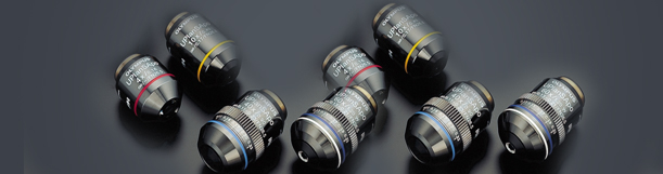
UIS Objectives For Life Science - The UIS objectives for life science include a wide range of top-class objectives that provide unrivalled clarity and which are in full compliance with international standards. This brochure includes detailed information on each series of available objectives, the applications and techniques they are best suited for, and an extensive objective performance chart.
Related Courses
· Live Blood Analysis (3 days)
· Live Blood Analysis (6 days)
· Live Blood Analysis (80 hours)
· Live Blood Analysis online course (1year)
· Colon Hydrotherapy Course 45 hours (6 Days)
· Cleansing Combo Pack
· Microscope Equipment
For detailed information about the Olympus FluoViewTM microscopes and available support equipment for the innovative microscopist, the appropriate brochures may be downloaded in this section. Extensive product photographs, charts, descriptions, sample images, technical specifications, and application notes can help you decide upon the perfect microscope, imaging system, software, and accessories to meet your specific research needs. Included in this section are brochures on research microscopes, digital camera systems, laser systems, objectives, dichromatic mirror units, software, and research microscopes.
FluoViewTM FV1000 Confocal Laser Scanning Biological Microscope - A leader in the next generation of live cell confocal imaging systems, the FV1000 features simultaneous laser light stimulation and imaging by incorporating two laser scanners in a single compact design. Synchronization of these two functions ensures that cellular reactions occurring immediately following or during stimulation are not overlooked, a key feature in microscopes designed for FRAP, FLIP, and photo activation. This 12-page full color brochure introduces the latest Olympus confocal instrument and reviews specifications, accessories, and configurations.
FluoViewTM Confocal Laser Scanning Biological Microscopes - Covering the entire Olympus FluoViewTM lineup of confocal microscopes, this 19-page full color brochure reviews the features currently available, including system components, specifications, objectives, scanning units, lasers, wavelength selection, software, three-dimensional imaging, and specialized techniques. In addition, a wide-ranging applications gallery is included to demonstrate instrument performance in real-world biological investigations, and a chart of fluorescent dyes can be used as a reference in choosing the appropriate fluorophores for your investigations.
AX70 Research System Microscope - By providing all of the advantages of the proprietary UIS infinity-corrected optical system, which provides the best image quality possible, as well as universal adaptability for rapid changeovers between various scientific techniques, a specially designed frame that ensures user comfort, and a choice of illuminator systems and accessories, the Olympus AX70 ranks as one of the most versatile research microscopes on the market today. Also, when coupled with its automatic photomicrography system, the AX70 offers a very advanced level of microscope performance and sophisticated photomicrography capabilities.
AX70-Macro Research System Microscope - The AX70-Macro is an advanced research system microscope whose wide field of view and use of a 0.5x objective allow observation of specimens up to 53 millimeters in diameter. Capable of brightfield micro and macro observations, the instrument is suitable for a wide range of applications, including the examination of large specimens, such as complete brain slices or small animals. Changing between observation types is easily accomplished by means of controls placed at the operator's hand.
AX80 Automatic Research Photomicroscope - A state-of-the-art microscope, the Olympus AX80 offers unprecedented levels of automation and precision to fulfill the most demanding requirements of cutting-edge research. The sophisticated instrument is outfitted with world-class optics, a revolutionary auto focus system, a simplified method changeover system, a universal photo system, and a vibration-resistant frame. The AX80 also can easily facilitate complex procedures involved with fluorescence in situ hybridization (FISH), patch clamping, and other biological research applications when its multi control box is linked to an external computer via an interface.
Blue Diode Lasers for Confocal Microscopy - Introducing the stable, high-performance 405-nanometer diode laser, this brochure describes how the system is modulated to permit high speed control of excitation light intensity over a selected region of the specimen. The document also reviews accompanying software and water immersion objectives corrected for spherical and chromatic aberrations in the visible light region. Detailed specifications for the laser system are also presented.
BX51 System Microscope (BX2 Series) - The Olympus BX51 universal research microscope features superb optical performance with a wide variety of contrast enhancing techniques, such as fluorescence, differential interference contrast, phase contrast, and darkfield. Built around a structurally rigid and compact Y-shaped motif, the BX51 is an excellent research-level imaging platform exhibiting versatility, a wide range of accessories, logical layout, and an ergonomic design.
BX51/BX61 Motorized System Microscope (BX2 Series) - The Olympus BX51 and BX61 microscopes offer top-quality optics and a full range of motorized modules that can be combined as the user requires, providing a level of performance that is more functional, more versatile, and more adaptable to the needs of advanced applications, such as the acquisition of multi-labeled fluorescence images, telemicroscopy, confocal microscopy, and automatic capturing of time lapse images. Selective addition of only the desired accessories allows maximum flexibility for automated imaging solutions.
BX51WI/BX61WI Fixed Stage Upright Microscope (BX2 Series) - Designed to achieve an even higher standard of stability and reliability in electro-physiological applications, the Olympus BX51WI and BX61WI fixed stage microscopes are equipped with a wide range of advanced new features to avoid vibration, including an intermediate magnification changer that can alter the magnification without changing objectives. Also, on top of the many benefits it shares with the BX51, the BX61 microscope incorporates a precise Z-axis focus motor into its durable frame.
BX52 Research Microscope (BX2 Series) - In addition to all of the advantages of the BX51, the Olympus BX52 offers rock-solid stability and rigidity enhanced by a special composite material of ceramic and aluminum, as well as superior fluorescence technology. Designed and built for leading research applications, the BX52 utilizes a newly-developed aspheric lens in the lamp housing to improve light collection and achromatic performance right up to near infrared conditions and meets all of the requirements for advanced laser microscopy, three-dimensional image analysis, and time-lapse experiments.
IX71 Research Inverted System Microscope (IX2 Series) - The Olympus IX71 microscope system features a rigid, compact frame, a two-tiered multi-port design, and outstanding UIS optical performance. The inverted instrument is also equipped with new fluorescence accessories and a V-shaped optical path for improved sensitivity in critical applications, while an external power supply ensures a stable platform for time-lapse observations. Other advanced applications that the IX71 facilitates include microinjection, multi dimensional imaging, and total internal reflection microscopy (TIRFM).
IX81 Motorized Inverted System Microscope (IX2 Series) - An extremely advanced motorized microscope, the Olympus IX81 provides high standards of observation, measurement, and functional operation. All major functions can be motorized, including focus, illumination, objectives, and optical path selection, and these motorized units may be operated via a remote handset, computer, or buttons on the microscope body. Additionally, the IX81 features numerous input and output ports, facilitating the use of several light sources and detectors.
DP12 Microscope Digital Camera - The Olympus DP12 digital camera provides high-resolution 3.34 million pixel imaging in a compact, easy-to-use design. The camera, which can be operated entirely from a control unit, is equipped with a universal C-mount thread that allows attachment to almost any microscope and a tiltable 3.5 inch LCD monitor that provides real-time display of large, easy-to-see images for faster, more accurate focusing and framing.
DP70 Digital Camera - Capable of capturing 12.5 million pixels in only 3 seconds, the Olympus DP70 enables users to quickly obtain accurate images with remarkable clarity, detail, and color depth. Coupled with advanced, multi-function software, the DP70 optimizes real-time acquisition conditions and subsequent image management, simplifying the strenuous demands of modern microscopy.
Q-Color 3 Imaging System - The Q-Color 3 digital imaging system delivers high resolution color images for microscopy documentation and publication. Included in the system are a 3.2 megapixel CCD sensor with a FireWire IEEE digital interface, QCapture software that is compatible with both Windows and Mac based systems, and Adobe Photoshop Elements for image enhancements. A Software Development Kit is also available for interfacing with custom software.
Q-Color 5 Imaging System - Similar to the Q-Color 3, the Olympus Q-Color 5 digital imaging system includes a CCD sensor and advanced acquisition and enhancement software. However, this state-of-the-art device is capable of even greater resolution with 5.0 active megapixels. Control of the camera is made simple through a single wire connection to a desktop or laptop computer.
MagnaFire/MagnaFire SP Biomedical Research Cameras - Compatible with Olympus MicroSuite software, both the MagnaFire and the MagnaFire SP are megapixel imaging camera systems designed for scientific microscope imaging applications. Some of the key features shared by the cameras are full resolution, real time focusing and framing via a FireWire interface, a live mode that ensures a flicker free image even at short exposures, Turbo gain control that reduces exposure duration and eliminates the need for binning, color merge, and real time image processing.
MicroFire Digital Camera - The Olympus MicroFire, a 2 megapixel FireWire digital color camera, acquires images with superb color fidelity and detail. Easy to install and use, this versatile camera offers high sensitivity, resolution, and dynamic range, making it an ideal choice for a wide array of microscopy applications. The camera even presents several solutions for fluorescence imaging when image signals are weak, smoothly facilitating one the most difficult imaging processes.
MicroSuite Basic - The MicroSuite Basic software package provides microscopists with the necessary tools to overcome common imaging problems. Through the use of an intuitive user-friendly interface, users can easily carry out image acquisition, multiple image alignment, image processing, database management, report generation, documentation, and movie creation.
MicroSuite B3 Biological Suite - Based on the analySIS software by Soft Imaging System, MicroSuite Biological Suite B3 is the natural choice for biological research. The package, designed for users with demanding biological applications, offers a fully automated measurement and analysis system. In addition, the B3 includes specialized image processing modules, including one for multiple fluorescence information processing, one for extended focal imaging, and one for multiple image alignment.
MicroSuite ScopeView Microscope Control Software - An invaluable extension to MicroSuite B3 and M3, ScopeView enables full computer control of all motorized components of the Olympus BX61, IX81, and AX70 microscopes. By facilitating such activities as motorized stage control, storage of easily recallable individual microscope configurations, and the linkage of microscope settings, this exciting software can save researchers both time and labor.
TIRFM Total Internal Reflection Fluorescence Microscope System - A technique especially useful in studies of cellular membrane function and single molecule events, total internal reflection microscopy is readily facilitated by the innovative Olympus TIRFM microscope system. The easily adjustable instrument may be equipped with two high numerical aperture objective lenses designed for evanescent wave illumination and up to three lasers can be introduced simultaneously with the optional laser combiner unit.
UIS Fluorescence Mirror Units - Designed to present the technical specifications of UIS fluorescence mirror units, this brochure offers a detailed unit selection chart based on fluorochrome characteristics. Mirror unit descriptions, a graphic representation of the characteristics of mirror unit wavelength, as well as information on custom fluorescence mirror units, fluorescence excitation balancers, and light source spectral characteristics are also provided.
UIS Objectives For Life Science - The UIS objectives for life science include a wide range of top-class objectives that provide unrivalled clarity and which are in full compliance with international standards. This brochure includes detailed information on each series of available objectives, the applications and techniques they are best suited for, and an extensive objective performance chart.
Related Courses
· Live Blood Analysis (3 days)
· Live Blood Analysis (6 days)
· Live Blood Analysis (80 hours)
· Live Blood Analysis online course (1year)
· Colon Hydrotherapy Course 45 hours (6 Days)
· Cleansing Combo Pack
· Microscope Equipment
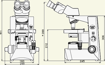
The CX31 microscopes improve all-round performance and offer excellent cost-efficiency.
 Advanced optical performance with excellent cost-efficiency.
The Olympus CX2 microscopes, which have gained an outstanding worldwide reputation in many medical and educational arenas, now evolve with new UIS2 infinity optics. The CX31 microscopes improve all-round performance and offer excellent cost-efficiency.
Outstanding flat images from PLCN objectives
The PLCN series of UIS2 objectives improves flatness dramatically, producing sharp, clear images right up to the edge of the field of view. Ideal for the 10X and 40X objectives so frequently used for inspection work, the flatness ranks among the very best in this class of microscopes.
Bright, even observation images
The illumination system employs a high intensity 6V, 30W halogen lamp for bright images. The aperture iris diaphragm with built-in condenser and standard field stop combine to provide bright and even illumination at all levels of magnification.
Rigid, durable construction for high performance and long service life
Construction quality is excellent throughout, with objectives, eyepiece,observation tube, revolving nosepiece, highly reliable rackless stage and other components all fixed firmly to the body — so there's nothing to come loose or fall off when the microscope is being transported. The highly reputable rackless stage is employed, and since the X-axis guide does not protrude, both transportation and operation are performed easily and safely.
For digital imaging (optional)
An optional adapter to mount Olympus digital cameras is provided to allow easy, costefficient digital imaging (sold separately).
Anti-fungus treatment
The treatment applied to the observation tubes, eyepieces and objectives, protects quality of optical parts even in high humidity regions
Related Courses
· Live Blood Analysis (3 days)
· Live Blood Analysis (6 days)
· Live Blood Analysis (80 hours)
· Live Blood Analysis online course (1year)
· Colon Hydrotherapy Course 45 hours (6 Days)
· Cleansing Combo Pack
· Microscope Equipment
Saturday, November 25, 2006
The Pacelli Chiropractic & Health Potential Complex Web site states:
How often do you get to see if small changes in nutrition, really can make a difference?
Well in this case, that's how we work. After making our recommendations, we repeat the test in 2 weeks, and look to see if we see changes.
This way you get to see the changes in your blood as we help correct the problems, it also allows us to check dosages to make sure that you are getting the proper amount of the vitamin, mineral, protein, or enzyme you need to restore you to optimal health.
These claims are sheer bunkum. Dark-field microscopy is a valid scientific tool in which special lighting is used to examine specimens of cells and tissues. The objects being viewed stand out against a dark background-the opposite of what occurs during regular microscopy.
This allows the observer to see things that might not be visible with standard lighting. Connecting a television monitor to a microscope for diagnostic purposes is also a legitimate practice. However, live cell analysis is not. Although a few characteristics of blood (such as the relative size of the red cells) are observable, live cell analysts invariably misinterpret other things, such as the extent of red blood cell clumping, changes in the shape of the cells, and other artifacts that occur as the blood sample dries.
Moreover, most practitioners who perform the test are not qualified to manage the problems they purport to diagnose.
During the mid-1980s, one company marketing live-cell equipment projected that a practitioner who persuades one patient per day to embrace a supplement program based on the test would net over $60,000 per year for testing and supplement sales. Another company estimated that with five new patients a day (22 days a month) paying $30 for the test and $50 for supplements, practitioners would gross over $100,000 per year just on initial visits.
Today's most active individual promoter of live-cell analysis is probably James R. Privitera, M.D., of Covina, California, who claims that "clot malfunction" is an underlying cause of many diseases, can be diagnosed with live cell analysis, and can be treated with large doses of dietary supplements.
His book, Silent Clots, describes his "general daily guidelines [for supplements] that have worked well for many patients as an anti-clotting program." The book also describes regimens for arthritis, asthma, baldness, bladder infections, cancer, colds, colitis, cramps, diabetes, diarrhea, diverticulosis, eczema, and edema, and includes case histories of patients he treated for many other conditions [1].
I do not believe there is any scientific evidence for these claims or that these regimens are effective as Privitera claims. His's web site offers more than 150 supplement products for sale.
In 1975, Privitera was convicted of conspiring to prescribe and distribute laetrile and was sentenced to six months in prison. (Laetrile is a quack cancer remedy.) In 1980, after the appeals process ended, he served 55 days in jail but was released after being pardoned by California Governor Jerry Brown. (The pardon occurred in response to a letter-writing campaign generated by the National Health Federation, a group that espouses what it calls "health freedom.")
Then, because Privitera had been prescribing unapproved substances (including laetrile, calcium pangamate, and DMSO) for the treatment of cancer, the California Board of Medical Quality Assurance suspended his medical license for four months and placed him on ten years' probation under board supervision.
During the probationary period, Privitera was "prohibited from making any representation that he is able to cure cancer through nutrition." He was also forbidden to tell patients they had cancer unless the diagnosis was confirmed in writing by an appropriate board-certified specialist.
During the probationary period, Privitera commercialized live-cell analysis and founded two companies that marketed devices for doing it. Silent Clots mentions that in 1993, a federal judge signed an order authorizing Internal Revenue Service agents to enter his clinic premises to effect a levy and that a seizure was made.
However, the book provides no further details about his tax-related difficulty.
Critical Reports
During the mid-1980s, National Council Against Health Fraud vice president James Lowell, Ph.D., watched three practitioners demonstrate live cell analysis at health expositions.
Lowell noted:
None took precautions during the preparation of the slides to prevent the blood from drying out or clotting.
They failed to clean their microscope slides carefully between patients, which meant that dirt seen under the microscope would be misinterpreted as blood components.
Some of the patterns one practitioner saw resulted from his microscope being out of focus and disappeared when Lowell adjusted it properly. [2}
Live cell analysis is also promoted by Infinity
2, of Scottsdale, Arizona, a multilevel company whose distributors often demonstrate their wares at chiropractic conventions. Infinity2 calls the test "live cell microscopy" and recruits licensed health professionals to use the procedure to sell its products. In 1995, I was tested at a national chiropractic convention where two teams of Infinity distributors had exhibits.
One exhibitor said I had "mild B12 deficiency" and "maldigestion" that could weaken my immune system and cause fatigue. The other said my blood cells showed evidence of "liver toxicity," "bacterial infection," and "free radical damage."
In both cases the recommended "treatment" was enzyme pills, which the company was marketing with false claims that "enzyme deficiency" is widespread among Americans. To reinforce their that the pills could help me, both practitioners gave me enzyme pills, repeated the procedure several minutes later, and said that the problems were no longer visible. Unknown to them, however,
I had faked taking the pills, so the "improvements" they saw had nothing to do with them. The most likely explanation is that the specimens were examined differently. Blood dries more quickly near the edges of the slide than near the center. Thus "improvement" will occur if the first specimen is examined near an edge and the second specimen is examined near its center.
Legal Action
In 1996, the Pennsylvania Department of Laboratories informed three Pennsylvania chiropractors that Infinity2's "Nutritional Blood Analysis" could not be used for diagnostic purposes unless they maintain a laboratory that has both state and federal certification for complex testing [3].
The company's attorney replied:
As background, you should know that part of our business activity is supplying a microscope system that is equipped for the demonstration of blood samples on a video monitor. We teach and recommend the use of this system as a health education demonstration. In our training and system we do not teach, promote or address in any manner using the system for "analysis" or any other medical purpose.
Our system is based on obtaining a written nutritional and lifestyle analysis from patients of doctors using the microscopy system. Once this questionnaire and evaluation of lifestyle and nutrition is examined by the doctor, the doctor then makes specific recommendations to the patient, based solely and exclusively on the nutritional analysis and personal interview with the patient.
After any recommendation has been made for altering the patient's lifestyle or nutritional habits, the doctor may place a single drop of blood on a sample slide for viewing through the dark field microscope which is then displayed on a video monitor.
This demonstration is a powerful motivating tool to encourage a doctor's patient to accept the doctor's previously made recommendations and alter his or her lifestyle to a healthier form of living.
The doctor performing the demonstration never makes any analysis, determination or recommendation based on the video demonstration. There is no commentary given regarding the state of the patient's blood or anything seen in the bloodscreen itself other than to educate the patient by pointing out features of the blood such as red cells, white cells, and plasma. The patient is able to view a sample of his/her own blood, which results in the motivation that can lead to beneficial lifestyle changes for the individual patient [4].
A Laboratory Department official then informed the attorney that certification would be required if the practitioner:
Either prescribed a nutritional supplement or changed the amount or type of a supplement based on viewing a blood sample.
Recommended lifestyle or nutritional changes based on viewing a blood sample.
Performed nutritional microscopy more than once for a given patients.
Compared the patient's blood smear to that of another slide or picture
Showed the patient's blood smear and making any comment or providing any information other than to point out features of the blood such as red cells, white cells, and platelets [5].
Very few if any chiropractors would have reason to seek laboratory certification, and even if they did, I doubt that permission would be granted to perform this test for diagnostic purposes. If you encounter anyone who performs any type of live cell analysis, please report this to your state department of laboratories and send a copy of your notice to me at P.O. Box 1646, Allentown, PA 18105
Reference
1. Privitera J, Stang A. Silent Clots: Life's Biggest Killers. Lockstep Medicine's Conspiracy to Suppress the Test That Should Be Done in Emergency Rooms Throughout The World. Covina, Calif.: The Catacombs Press, 1996.
2. Lowell JA. Live cell analysis: High-tech hokum. Nutrition Forum 3:81-85, 1986.
3. Wlazelek A. Chiropractors cease blood cell show and tell. State restricts the use of magnified images to sell vitamins, supplements. The Morning Call, April 12, 1996, page B6.
4. Woodruff SM. Letter to Joseph W. Gasiewski, Director, Division of Laboratory Improvement, Pennsylvania Department of Health, Jan 17, 1996.
5. Gasiewski JW. Letter to Samuel M. Woodruff, General Counsel, Infinity2, Feb 16, 1996.
Chirobase
The web site of the Integrated Medical Center, Annandale, Virginia, states:
 Blood is magnified by 30,000 times, giving us the ability to document your condition, monitor your progression while on treatment.
The blood is the pathway to the flesh and tells all. Using this technology allows us to monitor your condition and adjust as needed.
Your blood is the best place to find quick result and determine if you therapy is effective.
Quick response spells maximum results.
According to a flyer from a Los Angeles chiropractor:
 Live Blood Analysis is a simple procedure for obtaining a quick and accurate assessment of your blood.
With only a sample, taken virtually without pain from your finger, [the test] is able to provide a composite of over 25 aspects from your live blood.
Darkfield microscopy now allows us to observe multiple vitamin and mineral deficiencies, toxicity, tendencies toward allergic reaction, excess fat circulation, liver weakness and arteriosclerosis.
Live blood cell analysis
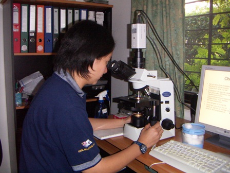
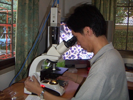 Live blood cell analysis is carried out by placing a drop of blood from the patient's fingertip on a microscope slide under a glass cover slip to keep it from drying out.
The slide is then viewed at high magnification with a dark-field microscope that forwards the image to a television monitor.
Both practitioner and patient can then see the blood cells, which appear as dark bodies outlined in white. The practitioner may take polaroid photographs of the television picture or may videotape the procedure for himself and/or the patient.
The results are then used as a basis for prescribing supplements. The procedure is also called live cell analysis, dark-field video analysis, nutritional blood analysis, libe blood analysis, and several other names. Most of its users are chiropractors, naturopaths, or bogus "nutrition consultants."
Related Courses
· Live Blood Analysis (3 days)
· Live Blood Analysis (6 days)
· Live Blood Analysis (80 hours)
· Live Blood Analysis online course (1year)
· Colon Hydrotherapy Course 45 hours (6 Days)
· Cleansing Combo Pack
· Microscope Equipment
Saturday, November 11, 2006
Wake Up to a new level of health.
 Wake Up to a new level of health. Wake up knowing you are healthier than you were yesterday.
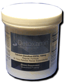
Detoxamin Ca-EDTA Chelation therapy works while you sleep to gently and gradually rid your body of heavy metals and other deadly toxins.
Be Aware that the average body holds 1,000% percent more lead today than 100 years ago simply because of the environment we live in.
Chelation therapy is a safe, effective, inexpensive alternative to drugs and surgeries.
Chelation therapy has been successfully used for decades to treat illnesses such as heart disease, strokes, diabetes, and removing unnecessary heavy metals like lead, mercury, even pesticides.
Protect Yourself and your family with the most effective self-administered Ca-EDTA therapy available.
Detoxamin Ca-EDTA chelation therapy is just as effective as traditional intravenous methods-without any needles, repeated office visits, or time consuming intravenous treatments. At about 30% of the traditional cost.
Learn More about Detoxamin or order now. Nothing to lose, only better health to gain.
More information: Chelation
more discussion: Forum
· Addiction Forum · Ask the Doctors Forum · Ayurveda Forum · Ayurvedic & Thai Herbs Forum · Colon Cleansing Forum · Dental Forum · Diabetes Forum · Diet Forum · General Cleansing Forum · Hepatitis A, B. C Forum · Integrated Medicine Forum · Live Blood Analysis Forum · Ozone-Oxygen-Forum · pH - Alkaline - Acidity Forum · Weight Loss Forum
Do I need Ca-EDTA chelation therapy?
 We find ourselves existing in a far more toxic and hostile environment than our bodies were designed to handle.
Experts have shown that almost every health problem-from learning disorders to cancer and heart disease-is aggravated by the approximate 1,000% increase in lead levels in our bones.
In 1999, it was reliably reported that hearts with some form of disease have 20,000 times more toxic heavy metals than healthy hearts.
"Human exposure to heavy metals has risen dramatically in the last 50 years as a result of an exponential increase in the use of heavy metals in industrial processes and products." says Maile Pouls, Ph.D (Townsend Letter for Doctors and Patients, July 1999).
A recently concluded "Body Burden" study by New York's Mt. Sinai Hospital and the Environmental Working Group was reviewed by University of Oregon Professor Joseph Thornton: "It shows the universality of chemical contamination of people's bodies," Thornton said. All the studies "confirm the general message that everybody in our society has these chemicals building up.
Some people have it worse than others, but everyone has it. No one is clean anymore." (From Being Careful Can't Keep Chemicals Out of Your Body, Miami Herald, February 1, 2003.)
Today we know that about one out of every 2.5 Americans will get cancer. Ninety eight percent of cancer is caused by toxic chemicals. When 50% of all men and 33% of all women living now will die of cancer, something is terribly wrong. (Mortality from cancer was reduced by 90% during an 18-year study of 59 patients treated with Calcium-EDTA.
Visit our website to read this study, and over 40 others proving the efficacy of Ca-EDTA chelation therapy and Detoxamin.) We will all function better and live longer if we lower the overall burden of toxic metals within ourselves. If you eat or breathe, you will probably benefit greatly from chelation therapy.
more information: Chelation
more discussion: Forum
· Addiction Forum · Ask the Doctors Forum · Ayurveda Forum · Ayurvedic & Thai Herbs Forum · Colon Cleansing Forum · Dental Forum · Diabetes Forum · Diet Forum · General Cleansing Forum · Hepatitis A, B. C Forum · Integrated Medicine Forum · Live Blood Analysis Forum · Ozone-Oxygen-Forum · pH - Alkaline - Acidity Forum · Weight Loss Forum
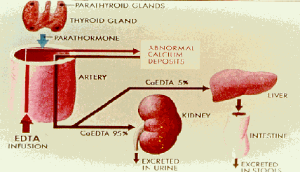
Answers to Some Important Questions about Ca-EDTA chelation therapy and Detoxamin. What is EDTA chelation therapy, and what is it used for?
 Chelation (pronounced key-lay-shun) is the process by which a metal or mineral (such as lead, mercury, iron, arsenic, aluminum, etc.) is bonded to another substance-in this case an amino acid called EDTA.
It is a natural process, basic to life itself. During EDTA chelation therapy, the EDTA infusion bonds with unwanted metals in the body and quickly carries them away in the urine.
Chelation therapy is a safe, effective alternative to drugs and surgeries and is used to treat many illnesses now known to be linked to the presence of toxic heavy metals.
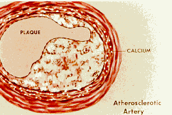
Illnesses such as heart disease, strokes, diabetes, circulatory disorders, neuropathies, Alzheimer's disease, atherosclerosis, and adverse reactions to many environmental pollutants.
Traditional chelation therapy uses an intravenous drip, and is administered in the outpatient setting.
The number of treatments vary based on each person's individual condition and/or goals of treatment. The average therapy is given one to three times a week for twenty to thirty treatments.
more information about Chelation
more discussion: Forum
· Addiction Forum · Ask the Doctors Forum · Ayurveda Forum · Ayurvedic & Thai Herbs Forum · Colon Cleansing Forum · Dental Forum · Diabetes Forum · Diet Forum · General Cleansing Forum · Hepatitis A, B. C Forum · Integrated Medicine Forum · Live Blood Analysis Forum · Ozone-Oxygen-Forum · pH - Alkaline - Acidity Forum · Weight Loss Forum
Detoxamin - New Patented Method of Chelation Therapy & EDTA Produces Superior Chelation Therapy Results
 Chelation and chelation therapy uses EDTA, a synthetic amino acid, and has been helping people with heavy metal toxicosis, heart disease and many other circulatory diseases for decades.
Chelation removes harmful toxins from your body, and because toxins are so pervasive in our food, air and water supply, anyone reading this now has a toxic metal burden.
Lead poisoning, mercury poisoning, arsenic poisoning in addition to many more toxins are wreaking havoc on our health. Detoxification for lead poisoning symptoms, mercury poisoning symptoms, and arsenic poisoning symptoms is accomplished successfully with the use of Detoxamin, the only patented method of chelation therapy in the world.
Tuesday, November 07, 2006
Two Prostate Cancer Radiotherapies Go Head-to-Head
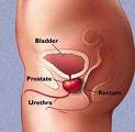 (HealthDay News) -- Prostate cancer patients treated with intensity-modulated radiation therapy (IMRT) suffer fewer long-term gastrointestinal side effects than those treated with another form of radiotherapy, new research shows.
Researchers at Fox Chase Cancer Center in Philadelphia analyzed data on 489 men treated with IMRT and 928 men treated with the second type of radiation therapy, called three-dimensional conformal radiation therapy (3D CRT).
The researchers evaluated gastrointestinal side effects such as short-term diarrhea and longer-term bowel dysfunction. They also looked at genitourinary side effects such as urinary frequency, urgency, painful or difficult urination, or symptoms of urinary obstruction.
"There were no differences in the reporting of acute gastrointestinal or genitourinary side effects for the two treatment modalities," study author Dr. Alexander Kirichenko said in a prepared statement.
"However, as the data are beginning to mature, we're seeing more long-term gastrointestinal side effects in the men treated with 3D CRT," Kirichenko added.
The finding is especially interesting because patients treated with IMRT receive higher radiation doses than those exposed to 3D CRT.
However, three years after treatment, gastrointestinal side effects were noted in 10.4 percent of patients treated with 3D CRT and in 6.3 percent of those treated with IMRT.
"Despite the specific findings pertaining to 3D CRT technique and the gastrointestinal side effects, men treated with either [technique] have acceptable rates of side effects at this point in our analysis, particularly when compared to data from surgical outcomes," Kirichenko said.
The findings were expected to be presented Sunday at the annual meeting of the American Society for Therapeutic Radiology and Oncology.
Both IMRT and 3D CRT enable doctors to precisely target cancer with multiple radiation beams, but IMRT allows doctors to control radiation dose intensity with far smaller radiation beams.
More information
The American Academy of Family Physicians outlines prostate cancer treatments.
Hot Air Spells Death for Head Lice
(HealthDay News) -- A contraption that looks like a cross between a vacuum cleaner and a hair dryer could rescue children from the scourge of head lice, a new study claims.
According to one of its creators, the device has a near-perfect success rate at killing off both lice and any of their eggs lurking in kids' hair. And the little critters shouldn't become immune to the so-called "LouseBuster," as they already have to some pesticides.
"It's extremely effective and extremely safe, and we think evolution-proof," said study co-author Dale Clayton, a University of Utah biology professor. "It would be very hard for insects to develop resistance to this assault."
According to Clayton, an estimated one in four American children will get infected by head lice. The tiny insects -- about the size of a sesame seed -- can be very difficult to eradicate.
One way is to get rid of them is to use repeatedly use special lice combs on children's heads, but this approach is so time-consuming that it can overwhelm parents. A variety of anti-lice shampoos are also available, but some parents don't like the idea of using insecticides -- including Malathion -- on their kids. Also, the U.S. Centers for Disease Control and Prevention says some lice have developed immunity to the chemicals used to kill them, although such problems are scattered.
Enter hot air, which some specialists think may be better at killing lice and their eggs.
Clayton and his colleagues tested a variety of hair dryers -- including handheld and "bonnet" models -- on 169 local children who were infested with lice. Their findings appear in the November issue of Pediatrics.
All the hair dryers killed at least 89 percent of lice eggs. But only one -- the specially designed "LouseBuster" -- managed to both kill eggs (98 percent) and wipe out high numbers of living lice (80 percent). The remaining living lice appeared unable to breed, perhaps due to stress or sterilization, the team said.
So, according to the study, the heads of children treated with the LouseBuster were free of lice one week after the half-hour treatment.
"We think it has a delayed effect on the lice it doesn't kill," Clayton said. "When you go back a week later, there's nothing there."
The air produced by the LouseBuster is hot -- much warmer than a typical hair dryer. Also unlike a hair dryer, it has a special handpiece designed to expose the roots of the hair.
The device apparently works by drying out the lice and their eggs, not by heating them, Clayton said.
The cost of the device is unknown, although Clayton estimated it should be in the hundreds of dollars, not the thousands, making it affordable for school districts. He predicted it could be on the market within a year or two, and added that the time required for treatment could eventually shrink to 15 minutes.
Dr. Craig Burkhart, a dermatologist at the Medical University of Ohio who studies lice, doubted that the device will be a success, however.
"The problem with the treatment is that it takes a half an hour at least to destroy the lice and the contraption is somewhat expensive and very cumbersome," he said.
What to do? "As with all bugs, insecticides remain the treatment of choice," Burkhart said.
More information
There's more on head lice at the U.S. Centers for Disease Control and Prevention.
Electric Current During Sleep Boosts Memory
 (HealthDay News) -- A popular theory suggests that sleep helps "fix" memories in the brain, and new research finds that passing a gentle electric current through the sleeping brain improves memory even more.
In this new study, researchers show that using an electric current at a particular frequency during non-rapid-eye-movement (non-REM) sleep can enhance memory about 8 percent.
"Students we tested were better at remembering a vocabulary list with oscillating electric stimulation," said lead researcher Jan Born, from the Department of Neuroendocrinology at the University of Luebeck, in Germany. "Between stimulation we observed increased slow oscillating activity, which is generated by the brain itself. This intensifies slow-wave sleep," he said.
His team reported the findings in the Nov. 5 online edition of Nature.
Intensifying slow-wave sleep enhances memory consolidation, Born said. Electric stimulation intensifies the brain's own slow oscillating activity, which drives the replay of recent memories. "This replay is the way memory is enhanced and retained," Born said.
In the study, Born's team applied electric stimulation to the scalps of 13 medical students who had been given a list of words to memorize before sleeping. While the students slept, five jolts of the current -- oscillating at frequencies similar to those seen naturally in slow-wave sleep -- were given over half an hour.
The researchers found that, at that frequency, students had an 8 percent better memory of the words on the list. However, if the frequency of the current or the phase of sleep in which it was given was changed, there was no improvement in memory.
It is possible that electric stimulation might enhance the memory of older patients or patients with Alzheimer's, Born said. "We can improve the function of slow-wave sleep and therefore memory," Born said. "It could also have an application in sleep disturbances, because this stimulation also improves sleep."
One expert thinks that these findings add to the evidence that sleep is involved in memory and learning.
"This is an interesting article that buttresses the important argument that sleep is an active, not a passive, state," said Dr. Robert D. Vorona, an associate professor in the Division of Sleep Medicine at Eastern Virginia Medical School. "We are beginning to understand that specific stages of sleep and regions of the brain likely play a role in different types of learning."
In addition, it raises the question as to whether one might eventually be able to alter the electrical and chemical aspects of sleep to optimize learning, Vorona said. "For now, however, both adequate amounts of study and sleep are important for learning," he said.
Another expert isn't sure that memory and sleep are connected.
"I am skeptical about sleep-learning studies in general," said Jerome M. Siegel, a professor of psychiatry and member of the Brain Research Institute at the University of California Los Angeles Medical Center. "This is a popular idea now, but it doesn't fit with a lot of other data."
Siegel noted that sleep-deprivation studies have shown that lack of sleep has no effect on memory of things such as word lists, names or events. "In sleep, brain metabolism is very low," Siegel said. "The idea that the brain is constructing memories at a time of reduced metabolism is not very compelling."
In cross-species studies, Siegel has also found there is no correlation between learning and the amount of sleep. "The animal that sleeps the most is the bat; the animal that sleeps the least is the elephant; I don't think that people think that bats are more intelligent than elephants," he said.
And no correlation between sleep and intelligence has been found in human studies, either, Siegel added.
"If you look at human sleep, in relation to other animals, both total sleep and REM sleep, the percent of sleep devoted to REM sleep is not unusual," he said. "If you are egotistical enough to believe that humans are the smartest species, it sure doesn't show in our sleep."
Sleep may be involved in memory consolidation, Siegel said, but it is not essential to strong memories.
More information
Find out more about sleep at the National Sleep Foundation.
Infant Iron Deficiency Lowers Teen Test Scores
 (HealthDay News) -- Teens who were iron-deficient as infants scored lower on cognitive tests of thinking, learning and memory than their peers, and this gap is particularly evident among poor children, new research shows.
Previous research has found that children with low levels of iron in their blood have worse scores on cognitive tests than those who aren't iron-deficient. Many children around the world have low iron levels.
In this study, University of Michigan researchers studied 185 children who lived in an urban area of Costa Rica. The children were an average of 17 months old when they were first screened for iron deficiency when the study began in 1983 to 1985.
The children were given cognitive tests at the beginning of the study and again at ages 5, 11 to 14, 15 to 18 and 19 years.
Of the children in the study, 87 were from middle-class families and 98 were from poorer families. The researchers found that 62 percent of the children with chronic iron deficiency were from poorer families.
Among the children from middle-class families, initial scores on cognitive tests were 101.2 for those with iron deficiency and 109.3 for those with sufficient iron levels. This gap remained at eight or nine points until the children were 19 years old.
Among the children from poorer families, the initial scores on cognitive tests were 93.1 for those with iron deficiency and 102.8 for those with normal iron levels. By age 19, that gap had widened to 25 points -- 70.4 vs. 95.3.
That kind of difference is likely to have a major impact on success in school and career choices in adulthood, the study authors noted. They said the findings emphasize the need to identify children at risk for iron deficiency in order to take steps to prevent or treat the condition in infancy.
The findings were published in the November issue of the journal Archives of Pediatrics and Adolescent Medicine.
More information
The Nemours Foundation has more about children and iron.
Subscribe to:
Posts (Atom)
Advanced Body Cleansing Kit$147.75  Advanced Body Cleansing Kit with Livatrex™, Oxy-Powder®, Latero-Flora™ and two bottles of ParaTrex®. |